ORIGINAL ARTICLE - Case Report
CITATION: Niranjan G, Prasad P, Verma A, Kumar A. Cytological Diagnosis of Hepatic Metastasis from Rectal Gastrointestinal Stromal Tumor: A Case Report. Discoveries 2025; 13(1): e202. DOI: 10.15190/d.2025.1
Cytological Diagnosis of Hepatic Metastasis from Rectal Gastrointestinal Stromal Tumor: A Case Report
Gauri Niranjan 1, Pallavi Prasad 1, *, Archana Verma 1, Ashok Kumar 2
- 1 Department of Pathology, Sanjay Gandhi Postgraduate Institute of Medical Sciences, Lucknow, India.
- 2 Department of Surgical Gastroenterology, Sanjay Gandhi Postgraduate Institute of Medical Sciences, Lucknow, India.
* Corresponding author: Pallavi Prasad, Department of Pathology, Sanjay Gandhi Postgraduate Institute of Medical Sciences, Lucknow; Room no.-11, New Diagnostic Block, C-block, 1st floor, India; Email: pallavisgpgi@gmail.com
Abstract
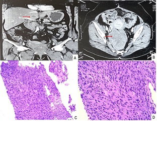
Gastrointestinal stromal tumours (GIST) are rare mesenchymal tumours which represent 1% to 3% of all gastrointestinal neoplasms. Rectal location of GIST is extremely rare accounting for 5% of GIST and only 0.1% of rectal tumours. They usually metastasise to the liver (65%). We hereby report a case of rectal stromal tumour with hepatic metastasis. A 55-year-old female presented with pelvic pain, associated with rectal bleeding. A thoracoabdominal computed tomography showed a large heterogeneous enhancing mass, arising from the rectum, anal canal and distal sigmoid colon measuring 12.3x8.7x7.6cm. Based on histopathological examination followed by immunohistochemistry, she was diagnosed with locally advanced rectal GIST. The tumour reduced in size after neoadjuvant-targeted treatment with imatinib. A local resection of the rectal GIST was successfully performed, and a diversion colostomy was done, later colostomy bag was attached. Following the operation, oral imatinib treatment was continued. On subsequent follow-up, her triple phase CECT whole abdomen showed multiple small well-defined peripherally enhancing hypodense liver lesions, the largest measuring 29x18mm suggestive of metastases. Ultrasound-guided fine needle aspiration from a liver lesion was reported as metastatic GIST. The patient underwent surgery, sunitinib was started and was discharged in stable condition. Thus, cytologic examination provides rapid interpretation, is a less invasive technique than open biopsy, and provides a cost-effective modality for diagnosing and managing inaccessible lesions.
Access FULL text of the manuscript here: FULL ARTICLE (PDF)
REFERENCES
1. Wu CE, Tzen CY, Wang SY, Yeh CN. Clinical Diagnosis of Gastrointestinal Stromal Tumor (GIST): From the Molecular Genetic Point of View. Cancers (Basel). 2019;11(5):679.
2. Turley RS, Peng PD, Reddy SK, Barbas AS, Geller DA, Marsh JW et al. Hepatic resection for metastatic gastrointestinal stromal tumors in the tyrosine kinase inhibitor era. Cancer. 2012;118(14):3571-8.
3. Søreide K, Sandvik OM, Søreide JA, Giljaca V, Jureckova A, Bulusu VR. Global epidemiology of gastrointestinal stromal tumours (GIST): A systematic review of population-based cohort studies. Cancer Epidemiol. 2016;40:39-46.
4. Miettinen M, Majidi M, Lasota J. Pathology and diagnostic criteria of gastrointestinal stromal tumors (GISTs): a review. Eur J Cancer. 2002;38 Suppl 5:S39-51.
5. Steigen SE, Eide TJ. Trends in incidence and survival of mesenchymal neoplasm of the digestive tract within a defined population of northern Norway. APMIS. 2006;114(3):192-200.
6. Tran T, Davila JA, El-Serag HB. The epidemiology of malignant gastrointestinal stromal tumors: an analysis of 1,458 cases from 1992 to 2000. Am J Gastroenterol. 2005;100(1):162-8.
7. Vij M, Agrawal V, Kumar A, Pandey R. Gastrointestinal stromal tumors: a clinicopathological and immunohistochemical study of 121 cases. Indian J Gastroenterol. 2010 Nov;29(6):231-6.
8. Vij M, Agrawal V, Pandey R. Malignant extra-gastrointestinal stromal tumor of the pancreas. A case report and review of literature. JOP 2011; 12: 200-204.
9. Fletcher CD, Berman JJ, Corless C, Gorstein F, Lasota J, Longley BJ et al. Diagnosis of gastrointestinal stromal tumors: A consensus approach. Hum Pathol. 2002;33(5):459-65.
10. Miettinen M, Lasota J. Gastrointestinal stromal tumors: pathology and prognosis at different sites. Semin Diagn Pathol. 2006;23(2):70-83.
11. Bamboat ZM, Dematteo RP. Updates on the management of gastrointestinal stromal tumors. Surg Oncol Clin N Am. 2012;21(2):301-16.12.
12. Yasui M, Tsujinaka T, Mori M, Takahashi T, Nakashima Y, Nishida T; Kinki GIST Study Group. Characteristics and prognosis of rectal gastrointestinal stromal tumors: an analysis of registry data. Surg Today. 2017;47(10):1188-1194.
13. Seidal T, Edvardsson H. Diagnosis of gastrointestinal stromal tumor by fine-needle aspiration biopsy: a cytological and immunocytochemical study. Diagn Cytopathol. 2000;23(6):397-401.
14. Li SQ, O'Leary TJ, Buchner SB, Przygodzki RM, Sobin LH, Erozan YS, Rosenthal DL. Fine needle aspiration of gastrointestinal stromal tumors. Acta Cytol. 2001;45(1):9-17.
15. Wieczorek TJ, Faquin WC, Rubin BP, Cibas ES. Cytologic diagnosis of gastrointestinal stromal tumor with emphasis on the differential diagnosis with leiomyosarcoma. Cancer. 2001;93(4):276-87.
16. Conrad R; ShobhaCastelino-Prabhu; Cobb C, Raza A. Role of cytopathology in the diagnosis and management of gastrointestinal tract cancers. J Gastrointest Oncol. 2012;3(3):285-98.
17. Inoue A, Ota S, Yamasaki M, Batsaikhan B, Furukawa A, Watanabe Y. Gastrointestinal stromal tumors: a comprehensive radiological review. Jpn J Radiol. 2022 ;40(11):1105-1120.
18. Lau S, Tam KF, Kam CK, Lui CY, Siu CW, Lam HS, Mak KL. Imaging of gastrointestinal stromal tumour (GIST). Clin Radiol. 2004;59(6):487-98.
19. Demetri GD, von Mehren M, Antonescu CR, DeMatteo RP, Ganjoo KN, Maki RG, Pisters PW et al. NCCN Task Force report: update on the management of patients with gastrointestinal stromal tumors. J Natl Compr Canc Netw. 2010;8 Suppl 2(0 2):S1-41; quiz S42-4.
20. Changchien CR, Wu MC, Tasi WS, Tang R, Chiang JM, Chen JS et al. Evaluation of prognosis for malignant rectal gastrointestinal stromal tumor by clinical parameters and immunohistochemical staining. Dis Colon Rectum. 2004;47(11):1922-9.